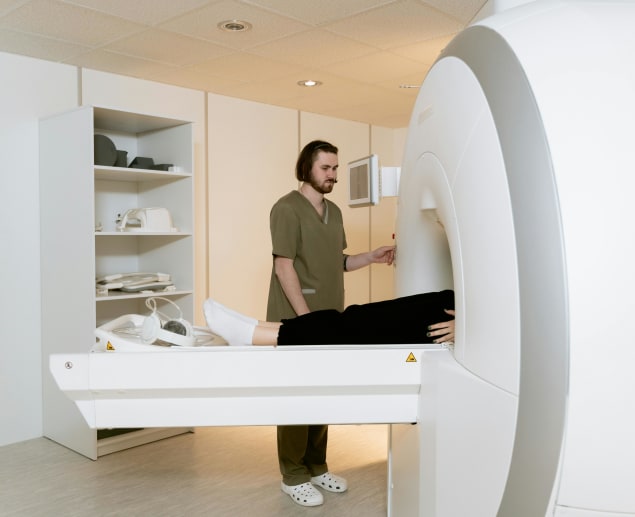
If you have concerns about ovarian cysts, understanding the right diagnostic tools can make all the difference. An MRI, known for its precision and detail, is a highly recommended imaging test that can be used to provide your healthcare provider with a clear picture of what’s happening inside your body.
In this comprehensive guide, we explore how an MRI ovarian cyst scan can identify an ovarian cyst and what type it is, determining whether it is harmless or potentially cancerous, and guiding effective treatment decisions.
What is an ovarian cyst?
An ovarian cyst is a fluid-filled sac that develops on or within an ovary
Ovaries are two small, almond-shaped organs positioned on either side of your uterus (womb) in your lower abdomen. Once a month, one of your ovaries releases a mature egg during ovulation. The ovaries also produce hormones oestrogen and progesterone that control your menstrual cycle and pregnancy.
Ovarian cysts are quite common, occurring naturally due to cycle-related changes during puberty, childbearing years, or menopause. Not at all cysts, however, are related to the menstrual cycle. They often do not cause any problems or symptoms and usually resolve on their own within a few weeks to months.
Ovarian cysts are mostly benign (noncancerous). But, there are instances when they are malignant (cancerous) or grow quite large, causing painful symptoms and leading to serious complications if left untreated.
Is an ovarian cyst the same as PCOS?
No, an ovarian cyst is not the same as PCOS.
Polycystic ovarian syndrome (PCOS) is a condition where you have multiple fluid-filled sacs surrounding your eggs, but these are actually called follicles, not cysts.
PCOS is a metabolic disorder that’s characterised by excess androgen (male sex hormone) production, leading to hormonal imbalance, excessive hair growth (hirsutism), irregular or absent periods, obesity, and fertility problems.
In contrast, an ovarian cyst is a fluid-filled sac on or inside an ovary. Unlike PCOS, an ovarian cyst can be asymptomatic, may disappear on its own, and does not typically disrupt the menstrual cycle.
What are the symptoms of an ovarian cyst?
As we mentioned earlier, ovarian cysts don’t cause any symptoms unless they persist and become very large. In that case, symptoms may include:
-
Pelvic pain – a dull, sudden, or sharp ache in the lower abdomen on the side of the cyst
-
A feeling of fullness or pressure (heaviness) in your abdomen
-
Bloating or abdominal swelling
-
Pain during or after sex
-
Loss of appetite or feeling full after eating a little
-
Problems with the menstrual cycle, such as irregular periods, heavier or lighter periods, spotting (abnormal bleeding between periods), or dysmenorrhea (painful periods)
-
An urgent or frequent need to urinate, pain during urination, or difficulty emptying the bladder (urinary retention)
-
Painful bowel movements
It’s important to seek medical advice or an MRI if you experience these symptoms, as they can cause complications.
Rarely, an ovarian cyst may rupture and bleed into the abdomen, increasing the risk of infections and creating a medical emergency.
A complication known as ovarian torsion can also occur when an ovarian cyst becomes so large that it causes the affected ovary to twist and knot around the ligaments that hold it in place, restricting blood flow and ultimately leading to its death.
Why do I need an MRI for ovarian cyst diagnosis?
A pelvic or transvaginal ultrasound scan is usually the first port of call when ovarian cysts are suspected, following a medical history review and thorough pelvic exam.
This scan uses high-frequency sound waves to visualise ovarian abnormalities. However, it can sometimes provide unclear or indeterminate results, failing to identify the content and nature of an ovarian cyst, especially when a complex cyst is detected.
Moreover, it can't definitively distinguish between a benign (non-cancerous) and malignant (cancerous) mass. In any case, your healthcare provider might order an MRI ovarian cyst scan to get clearer images and a more detailed analysis for a more accurate diagnosis.
A magnetic resonance imaging (MRI) scan is an imaging test that uses a strong magnetic field and radio waves to generate three-dimensional (3D), detailed images of the body’s internal structures, including bones, organs, and soft tissues such as muscles, blood vessels, ligaments, nerves, and more.
Here are some of the advantages of an MRI scan for ovarian cyst:
-
It is non-invasive.
-
It doesn’t involve exposure to radiation.
-
It offers excellent soft tissue contrast. This means that it shows distinct differences between various types of soft tissues in the body.
-
It delivers comprehensive information.
With an ovarian cyst MRI scan, you will get high-resolution images that'll allow your healthcare provider to view the cyst in fine detail, including its exact location, size, shape, structure, composition, and relation to surrounding tissues. This level of imaging detail is crucial, especially for complex ovarian cysts.
MRI for complex ovarian cyst diagnosis
Complex ovarian cysts differ from simple cysts because they may contain thicker fluid, such as mucus or blood, and solid material or areas.
They may also feature walls dividing the cyst into multiple fluid-filled areas (septations) or have visible or prominent blood vessels, indicating a greater blood supply flowing to and within them.
Complex cysts are not as common as simple cysts, and they usually develop from abnormal cell behaviour (e.g., overgrowth of cells) inside or around the ovaries.
An example is a complex ovarian cyst called endometrioma, formed when cells like those of the endometrium (the tissue lining the uterus or womb) grow on the ovaries.
Endometriomas are rarely malignant (cancerous). Even so, there are other types of complex cysts with different disease processes, characteristics, and compositions.
Determining the precise nature of these cysts is crucial, as they can sometimes indicate cancer. An MRI for complex ovarian cysts is particularly useful in these cases because it can clearly show the external and internal structure of the cysts, helping to tell simple cysts from complex ones and determining whether they are benign (non-cancerous) or potentially malignant (cancerous).
What does an ovarian cyst look like on an MRI?
On MRI ovarian cyst images, an ovarian cyst generally appears as a round or oval-shaped structure that is distinctly different from the surrounding healthy tissues.
Simple cysts typically look like clear, fluid-filled sacs and are usually uniform in appearance with smooth boundaries. Depending on the MRI techniques used, they are often bright or grey in the centre with a dark-coloured outline, making them stand out against the grey background of the surrounding ovarian tissue.
Complex ovarian cysts, on the other hand, contain both fluid-filled and solid areas, so they may appear heterogeneous (lacking uniformity) with internal structures or debris that vary in signal intensity (i.e., colour) and texture.
These internal structures may display shades of grey and white, reflecting their varying tissue types and densities. Complex cysts might also show irregular borders and septations (internal walls), which can give them a more complex appearance on images obtained from an ovarian cyst MRI scan.
Benign vs malignant ovarian cyst MRI
The difference between a benign and malignant ovarian cyst is simple.
- A benign cyst is non-cancerous. It may grow in size, but it doesn’t metastasize (spread to other body parts).
- Malignant ovarian cysts are cancerous and can spread to other organs like the liver, intestines, stomach and more. They typically manifest as solid tumours or complex cysts with solid areas, in contrast to simple ovarian cysts, which are fluid-filled masses.
MRI's advanced technology and range of techniques make it excellent for visualising the size and shape of a mass, but also its tissue characterisation. This means MRI can identify various tissue types inside an ovarian cyst, including fluid, fat, blood, mucus, or solid materials, to determine whether a lump is a cyst or a tumour.
With the comprehensive information obtained through MRI imaging of an ovarian mass, it becomes possible to determine whether an ovarian cyst is benign or malignant.
Can MRI detect ovarian cancer?
When abnormal growth is noticed anywhere in the body, many wonder about the possibility of cancer. So, while we’ve mentioned that ovarian cysts are mostly benign, you may be wondering if an MRI can detect ovarian cancer. The simple answer is yes!
An MRI ovarian cyst scan can distinguish between a non-cancerous (benign) and cancerous (malignant) ovarian mass, with an overall accuracy of 88% to 93% for the diagnosis of malignancy.
It also delivers visuals of surrounding tissues, such as the uterus and lymph nodes, making it easier to check whether the cancer has spread and to determine its current stage.
Sometimes, a gadolinium-based contrast agent or dye may be injected intravenously before or during an MRI scan to highlight certain characteristics that are indicative of malignancy, although contrast isn’t always necessary.
It is important to know that while an MRI is a powerful tool for ovarian cancer detection, it cannot definitively diagnose cancer—it can only suggest potential malignancy based on imaging characteristics. Further diagnostic tests, such as a targeted biopsy, will be required to confirm the presence of cancer.
How is an ovarian cyst treated?
Treatment for ovarian cysts varies based on your age, the type of cyst, its size, and the symptoms it causes. Here are some options your healthcare provider might suggest:
-
Watchful waiting - Most ovarian cysts are benign and resolve naturally over time. Therefore, the first line of action is usually to watch and wait, especially if the cyst is small, fluid-filled, and causing no symptoms. You will need to undergo several routine imaging tests to monitor the cyst's progress, whether it diminishes, enlarges, or changes in appearance. If your ovarian cyst is causing mild pelvic pain, you may be prescribed a nonsteroidal anti-inflammatory drug (NSAID) or advised to use over-the-counter painkillers while the cyst is being closely monitored.
-
Surgery - This treatment method is often employed for cysts that won’t go away or are very large, painful, and suspicious. The surgical procedure used to remove ovarian cysts is called cystectomy. It is performed in two different ways, namely:
-
Laparoscopy (keyhole surgery).
-
Laparotomy (open surgery) for very large cysts.
In some cases, an oophorectomy (removal of the ovarian cyst and the affected ovary) might need to be performed, depending on the size of the cyst, its nature, and whether it continues to recur.
What other scans can be used for ovarian cysts?
While an MRI for ovarian cyst scan can be trusted to provide detailed information, other scans can be used in its place. These include:
-
Ultrasound - This can either be an external pelvic or transvaginal (internal) scan. An ultrasound is the primary imaging tool when an ovarian cyst is suspected because it is highly effective in identifying the presence of a cyst and assessing its basic characteristics. Compared to other scans, it is quicker and relatively inexpensive.
-
Computed Tomography (CT) scan - A CT scan uses X-rays and a computer to visualise the body’s internal organs. It is particularly useful in situations where an MRI is contraindicated (i.e., not suitable for use). Contraindications for MRI include:
-
Presence of metallic foreign bodies or implants - The strong magnetic field used in MRI can interfere with metallic implants such as cardiac pacemakers, potentially affecting the quality of the imaging.
-
Pregnancy, serious kidney problems, or a known allergic reaction to contrast dyes - Detailed MRI ovarian cyst images can often be obtained without the use of a contrast agent, but some situations may require it for optimal imaging. This poses a risk to people who are pregnant, on dialysis or have previously experienced allergic or anaphylactic reactions to the gadolinium-based contrast used in MRI scans. Additionally, people who are diabetic or hypertensive might need to consult with a clinician before undergoing the procedure.
-
Claustrophobia or high body mass index (BMI) - Being claustrophobic or overweight can make it difficult or uncomfortable to fit into the narrow, enclosed space of an MRI scanner. An open and upright MRI scan can be used to address these issues and provide a more comfortable experience.
If you are unsure which imaging test is right for you, book a consultation for £50. An expert clinician will call you to discuss your medical history and offer a no-obligation referral to the scan best suited to your unique concerns.
Scan.com is the UK’s largest imaging network, with over 200+ scanning centres nationwide. Booking an MRI ovarian cyst scan, pelvic or transvaginal ultrasound or CT scan is as easy as browsing through locations near you, comparing prices, checking for the earliest appointment time, and hitting the ‘book now’ button. No GP referral needed. No NHS waiting lists.
Sources:
-
Benign Ovarian Cysts. (n.d.). Johns Hopkins Medicine. Retrieved May 18, 2024, from https://www.hopkinsmedicine.org/health/conditions-and-diseases/benign-ovarian-cysts
-
Ghadimi, M., & Sapra, A. (2023, May 1). Magnetic Resonance Imaging Contraindications - StatPearls. NCBI. Retrieved May 18, 2024, from https://www.ncbi.nlm.nih.gov/books/NBK551669/
-
Iyer, V. R., & Lee, S. I. (2010, February). MRI, CT, and PET/CT for Ovarian Cancer Detection and Adnexal Lesion Characterization. American Journal of Roentgenology. Retrieved May 21, 2024, from https://www.ajronline.org/doi/full/10.2214/AJR.09.3522
-
Kallen, A. (n.d.). Ovarian Cyst: Symptoms, Causes, Treatment and More. Healthline. Retrieved May 18, 2024, from https://www.healthline.com/health/ovarian-cysts
-
Mobeen, S., & Apostol, R. (n.d.). Ovarian Cyst - StatPearls. NCBI. Retrieved May 18, 2024, from https://www.ncbi.nlm.nih.gov/books/NBK560541/
-
Musheyev, Y., Levada, M., & Ilyaev, B. (2022, March). Ovarian Serous Cystadenoma Presents As Bladder Issues in 23-Year-Old Female: A Case Report. NCBI. Retrieved May 18, 2024, from https://www.ncbi.nlm.nih.gov/pmc/articles/PMC8910779/
-
Ohya, A., & Fujinaga, Y. (2022, August 2). Magnetic resonance imaging findings of cystic ovarian tumours: major differential diagnoses in five types frequently encountered in daily clinical practice. Japanese Journal of Radiology, 40, 1213–1234. https://doi.org/10.1007/s11604-022-01321-x
-
Ovarian Cystectomy: Purpose, Procedure, Risks & Recovery. (n.d.). Cleveland Clinic. Retrieved May 18, 2024, from https://my.clevelandclinic.org/health/treatments/24427-ovarian-cystectomy
-
Overview - - - Ovarian cyst. (n.d.). NHS. Retrieved May 18, 2024, from https://www.nhs.uk/conditions/ovarian-cyst/
-
Overview: Ovarian cysts - InformedHealth.org. (n.d.). NCBI. Retrieved May 18, 2024, from https://www.ncbi.nlm.nih.gov/books/NBK539572/
-
Park, S. B., & Lee, J. B. (2014, January). MRI features of ovarian cystic lesions. Wiley Online Library. Retrieved May 18, 2024, from https://onlinelibrary.wiley.com/doi/full/10.1002/jmri.24579
-
Puckett, Y., & Hoyle, A. T. (n.d.). Endometrioma - StatPearls. NCBI. Retrieved May 18, 2024, from https://www.ncbi.nlm.nih.gov/books/NBK559230/
-
Tests for Ovarian Cancer | How Is Ovarian Cancer Diagnosed? (n.d.). American Cancer Society. Retrieved May 18, 2024, from https://amp.cancer.org/cancer/types/ovarian-cancer/detection-diagnosis-staging/how-diagnosed.html
-
Valentini, A. L., Gui, B., Miccò, M., Mingote, M. C., Gaetano, A. M. D., Ninivaggi, V., & Bonomo, L. (2012). Benign and Suspicious Ovarian Masses—MR Imaging Criteria for Characterization: Pictorial Review. NCBI. Retrieved May 18, 2024, from https://www.ncbi.nlm.nih.gov/pmc/articles/PMC3321462/


