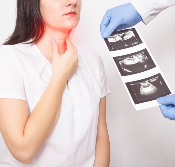

Thyroid Ultrasound: Information & Get a Scan
An ultrasound is a safe, painless way of diagnosing medical conditions that can affect various parts of the body. It uses high frequency sound waves to create images of the inside of the body that doctors can then use to determine if a disease or an abnormality is present.
A thyroid gland ultrasound focuses on the thyroid, a gland situated in the neck. It can help to diagnose conditions such as an under or an overactive thyroid, thyroiditis, goitres and thyroid cancer.
What is a Thyroid Gland Ultrasound?
The thyroid gland is a relatively small, butterfly-shaped gland that sits at the base of the neck, above the collarbones and in front of the windpipe, or trachea. It comprises a left and right lobe with a thin piece of thyroid tissue between them called the isthmus and is one of several glands that make up the endocrine system. The endocrine system is responsible for the manufacture of the hormones that regulate everything from mood and the sleep-wake cycle to blood pressure, blood sugar levels and body temperature.
Specifically, the thyroid gland is responsible for the hormones thyroxine and triiodothyronine that regulate metabolism, appetite and weight, and also play a role in regulating body temperature and blood pressure.
A thyroid gland ultrasound is a scan that looks at the health of the thyroid to help diagnose different types of thyroid disease.
Why a Doctor Might Order a Thyroid Ultrasound
A doctor may organise a thyroid ultrasound if they suspect you may be experiencing thyroid disease. Symptoms of a thyroid condition include unexplained weight loss or weight gain, tiredness and irritability. Some conditions that affect the thyroid tissue may also lead to tightness or a visible swelling in the neck.
What a Thyroid Ultrasound Can Diagnose
A thyroid gland ultrasound can be used to diagnose various conditions. Ultrasound imaging of the thyroid is often carried out alongside other tests including a physical exam where your doctor feels around your neck, and blood tests to test for thyroid function and thyroid hormone levels.
Your doctor may also perform a type of biopsy called a fine needle aspiration, or FNA, to remove a small piece of thyroid tissue or tissue from the nearby lymph nodes, for further examination, depending on your symptoms and medical history.
Thyroid ultrasounds can be useful in the diagnosis of the following conditions:
Thyroid Nodules
Thyroid nodules are small growths that can develop on the thyroid gland - they’re usually benign, but a thyroid nodule can become cancerous, leading to thyroid cancer. Most thyroid nodules are linked to an iodine deficiency.
Thyroid Cysts
Thyroid cysts are small fluid-filled growths that can develop on the thyroid gland. They’re usually benign, but if they grow larger, they can cause problems with breathing and swallowing, and may cause a tight feeling in the neck.
Goiter
A goiter is a non-cancerous, enlarged thyroid gland that leads to a swelling in the neck. This can then lead to difficulty breathing, swallowing and speaking. Goiters are usually caused by an iodine deficiency.
Thyroid Cancer
Sometimes, growths and swelling in the thyroid gland can become cancerous. A thyroid gland ultrasound, along with a fine needle aspiration biopsy, can help to diagnose or rule out thyroid cancer.
Thyroiditis
Thyroiditis describes an inflamed, swollen thyroid that can lead to hyperthyroidism or hypothyroidism (see below).
Graves’ Disease
Graves’ disease is the most common type of hyperthyroidism (see below). It’s a type of autoimmune disease and causes the immune system to mistake the thyroid gland for a foreign body and to attack it. This leads to anxiety, hand tremors, a racing heart, an increase in sweating, insomnia and weight loss.
Hashimoto’s Thyroiditis
Hashimoto’s thyroiditis is the most common form of thyroiditis and the most common cause of hypothyroidism (see below). It’s a type of autoimmune disease, where the thyroid tissue is mistakenly attacked by the immune system.
Hyperthyroidism
Hyperthyroidism is caused by an overactive thyroid that produces too much thyroid hormone. It’s characterised by weight loss, restlessness, heart palpitations, irritability, anxiety, sweating, thin skin, brittle nails and falling hair.
Hypothyroidism
Opposite to hyperthyroidism, hypothyroidism is caused by an underactive thyroid, and too little thyroid hormone production. It’s characterised by weight gain, depression, a slow heart rate, constipation, dry skin and hair, memory problems and an increased sensitivity to cold temperatures.
Lymph Node Enlargement
Sometimes, thyroid nodules, thyroiditis and thyroid cancer can lead to an enlargement of the thyroid gland and the nearby lymph nodes, leading to a visible swelling in the neck and difficulty breathing, swallowing and speaking.
Types of Thyroid Ultrasounds
There are different types of thyroid ultrasound - which one you have will depend on your symptoms. Your medical team will explain which type you’ll be having.
Conventional (Gray-Scale) Ultrasound
A conventional ultrasound is used to create images of the thyroid gland, along with the surrounding soft tissues, that appear in various shades of black, white and grey on the ultrasound monitor. It’s often the first test performed to ascertain if thyroid disease is present.
Doppler Ultrasound
A Doppler ultrasound is the same as a standard ultrasound, with extra capability used to measure blood supply. Your doctor may use this type of ultrasound, to help them differentiate between benign and cancerous nodules if abnormalities are seen on a conventional ultrasound. During a Doppler ultrasound, you’ll hear a whooshing sound as it picks up the sounds of the blood flow through the blood vessels.
Elastography
If abnormalities or signs of thyroid disease are detected, your doctor may also perform a thyroid elastography ultrasound to help them measure the stiffness of the thyroid tissue. This helps to differentiate between benign and cancerous thyroid nodules.
Contrast-Enhanced Ultrasound (CEUS)
A contrast-enhanced thyroid ultrasound uses a contrast dye, injected into a blood vessel, to help further highlight blood supply and to help differentiate between benign and malignant growths.
3D Ultrasound
A 3D thyroid gland ultrasound provides more detailed images of the thyroid, allowing a more comprehensive evaluation of the size, shape and structure of the gland itself, and any abnormalities.
How an Ultrasound of the Thyroid Works
An ultrasound uses sound waves to create images of the inside of the body. When used to scan the thyroid, a probe called a transducer is moved over the skin of the neck and throat, above where the thyroid gland is situated.
The transducer sends high frequency sound waves through the skin, helped by a special water-based conductor gel placed onto the skin. These sound waves then bounce off the internal organs, tissues, blood vessels and fluids and are sent back through the transducer to the ultrasound machine as an echo, that can then be turned into an image.
The high resolution ultrasound images can then be analysed by a specialist doctor called a radiologist who will then discuss them with your referring doctor.
Equipment Used
Ultrasound equipment includes the transducer and ultrasound machine, which are connected to a computer and a monitor that enables the radiographer to view the images.
Who Performs a Thyroid Gland Ultrasound?
A healthcare professional called a radiographer usually performs a thyroid gland ultrasound, usually in the radiology department of a hospital or specialist clinic.
Benefits
A thyroid gland ultrasound is a quick, painless test that allows doctors a clear view of the thyroid size shape and overall health - you’ll be able to return home the same day. It provides clear images of the soft tissues that can be difficult to achieve with an x-ray. Unlike x-rays and CT scans that carry a low risk for radiation exposure, an ultrasound doesn’t use radiation.
Ultrasounds of the thyroid can inform doctors where any growths or abnormalities may be, what size they are, if they have a blood supply and whether or not they’re filled with fluid. This data then helps doctors diagnose or rule out certain conditions.
Risks & Side Effects
There are no known risks or side effects to having an ultrasound scan - they’re considered safe, even for pregnant women. However, if you have an enlarged thyroid gland or you’re experiencing swelling or discomfort in your neck, it may be a little uncomfortable when the transducer is passed over the area.
Limitations
A thyroid ultrasound cannot give a diagnosis of an under or an overactive thyroid or some other types of thyroid disease alone. It needs to be carried out in conjunction with thyroid hormone blood tests and potentially a biopsy. Ultrasound results may also need to be assessed alongside results from an MRI scan to get a full diagnosis.
How to Prepare for a Thyroid Ultrasound
A thyroid ultrasound doesn’t require much preparation. When you arrive for your appointment, you will be asked to remove any jewelry or accessories from your neck area. You may be asked to remove your upper clothing and to wear a hospital gown.
You can eat and drink as normal beforehand, and your medical team will let you know if you should continue with any regular medications or if you should pause them, and if so, for how long.
The Procedure Explained: What to Expect
A thyroid ultrasound is a painless and relatively quick type of examination, usually taking around 30 minutes. You will be lying back on a hospital bed throughout, and you’ll need to tilt your head back to stretch your neck and throat to create more space for scanning.
Application of Gel on Neck
Your radiographer will apply a generous amount of conductor gel to your neck for the transducer to send (inaudible) high frequency sound waves and receive the echoes back.
Placement of Transducer on Thyroid Area
They will then place the transducer onto your neck area and slowly move it around from side to side, down towards your chest and up towards your head. If you have any discomfort or swelling, let them know and they will be as gentle as possible.
Real-Time Imaging and Assessment
The transducer will immediately start to pick up the echoes as the sound waves bounce off the thyroid gland and the surrounding tissues, blood vessels and lymph nodes. These echoes will then translate into images on the monitor, that are then seen in real-time.
Doppler Evaluation if Needed
If your medical team thinks they need information about the blood flow and health of the blood vessels feeding the thyroid, they will also perform a Doppler ultrasound. This will feel the same as a regular ultrasound, but you’ll hear a whooshing sound as the transducer picks up the sounds of the blood vessels.
Image Capture and Measurements
Your radiographer will pause the transducer in certain areas to capture the best images that are then saved to the computer in your medical notes. They will also use computer programming to measure the size and shape of the thyroid gland.
Removal of Gel
When your radiographer is happy that they have the right images and measurements, they will then use tissue to remove the gel from your skin. It may feel sticky afterwards, but it shouldn’t stain your clothing.
Review by Radiologist
Once the images are captured, they’ll be sent digitally to a radiologist who is trained to assess the differences between a healthy, normal thyroid, and one that is diseased, inflamed or not functioning properly.
What Happens After a Thyroid Gland Ultrasound?
After your radiographer is happy that they have sufficient images, they will stop the ultrasound and ask you to get dressed. They will then explain how long it might be before you get your results. You’ll be able to return home the same day.
Getting the Results
Your referring doctor will give you your results when they’ve spoken to the radiologist who reviewed them. They may also need to speak to other specialist doctors such as a specialist in endocrinology (hormones and the endocrine system) or surgeons if they think you may require surgery to your neck and thyroid gland. Your medical team will then discuss the next steps to get a definitive diagnosis of thyroid disease or to begin treatment.
Ultrasound Images
Ultrasounds provide detailed black, white and grey images of the soft tissues. The denser the tissue, the darker it will appear on an ultrasound.
Colours
If a Doppler ultrasound is performed, it will provide detailed information about the blood flow to and from the thyroid gland. Blood vessels that appear red indicate blood flowing towards the transducer, and blue blood vessels indicate blood flowing away from the transducer. The deeper the colour, the faster the blood flow. This can help doctors determine if thyroid cancer is present, and if a thyroid nodule or tumour has a blood flow to it.
Normal vs Abnormal
A normal thyroid will appear smooth with a uniform colour on an ultrasound, with each lobe symmetrical and a well-defined isthmus. Thyroid disease, be that due to benign nodules, cancerous tumours or any other abnormality will show as a larger, bumpy, inflamed or irregular-shaped thyroid gland. Radiologists and specialised doctors will know what signs to look out for when assessing and diagnosing thyroid disease.
Costs
A private thyroid gland ultrasound costs around £150 to £200 in the UK. Consultations and any treatment carried out privately will cost more.
Get a Thyroid Ultrasound
If you’re experiencing pain, discomfort, tightness or swelling in your neck, you’re having difficulty breathing, swallowing or speaking or you have unexplained weight loss, weight gain, tiredness, dry skin and hair, insomnia, a racing heart or excessive sweating, speak to your GP about having a thyroid gland ultrasound. You may have to wait several weeks or months via the NHS. If you’d like to book a private ultrasound, we can help get you an appointment as soon as possible.
FAQs
How Long Does a Thyroid Ultrasound Take?
A thyroid ultrasound usually takes around 30 minutes.
What Does Thyroid Cancer Look Like on an Ultrasound?
A cancerous thyroid gland will appear irregularly-shaped with visible nodules and increased blood flow.
What Is the Normal Size of the Thyroid Gland on an Ultrasound?
A normal thyroid gland in an adult is usually around 40-60mm wide.
References
Stang, D. (2020, February 27). Thyroid ultrasound. Healthline. https://www.healthline.com/health/thyroid-ultrasound
Website, N. (2025, February 26). Ultrasound scan. nhs.uk. https://www.nhs.uk/conditions/ultrasound-scan/
Thyroid Gland | North Bristol NHS Trust. (n.d.). https://www.nbt.nhs.uk/our-services/a-z-services/endocrine-surgery/thyroid-gland
Wallace, R. (2023, May 9). The 6 common thyroid Problems & Diseases. Healthline. https://www.healthline.com/health/common-thyroid-disorders
